Gynecologist Appointment
About
Having a continuous individual pregnancy monitoring by the same gynaecologist has been shown to be beneficial for satisfaction with medical care, as well as safety.
Our gynaecologist, midwives and other members of the interdisciplinary team work closely together, to achieve an individually tailored pregnancy care for you and your family.
The pregnancy monitoring by our gynaecologist, Dr. Liina Rajasalu, is performed in our partner clinic - Valvekliinik (Pärnu mnt 48a).
Some international insurances or private insurances cover theses costs.
Take a look at our FQA or contact us directly.
Packages
-
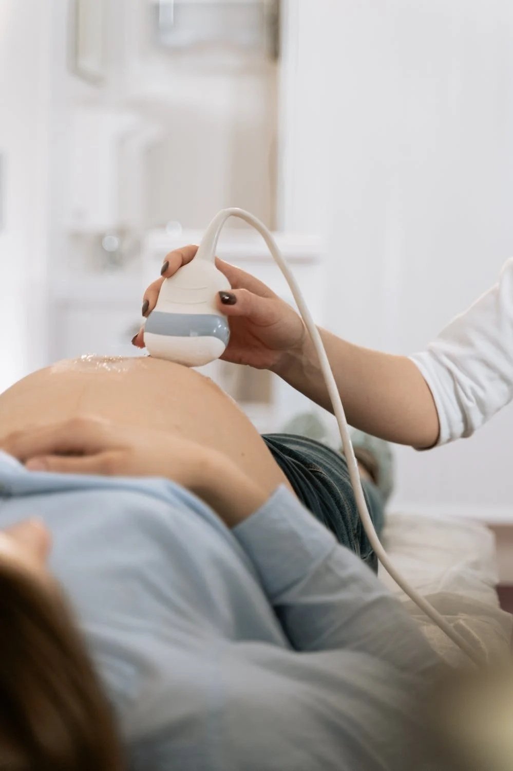
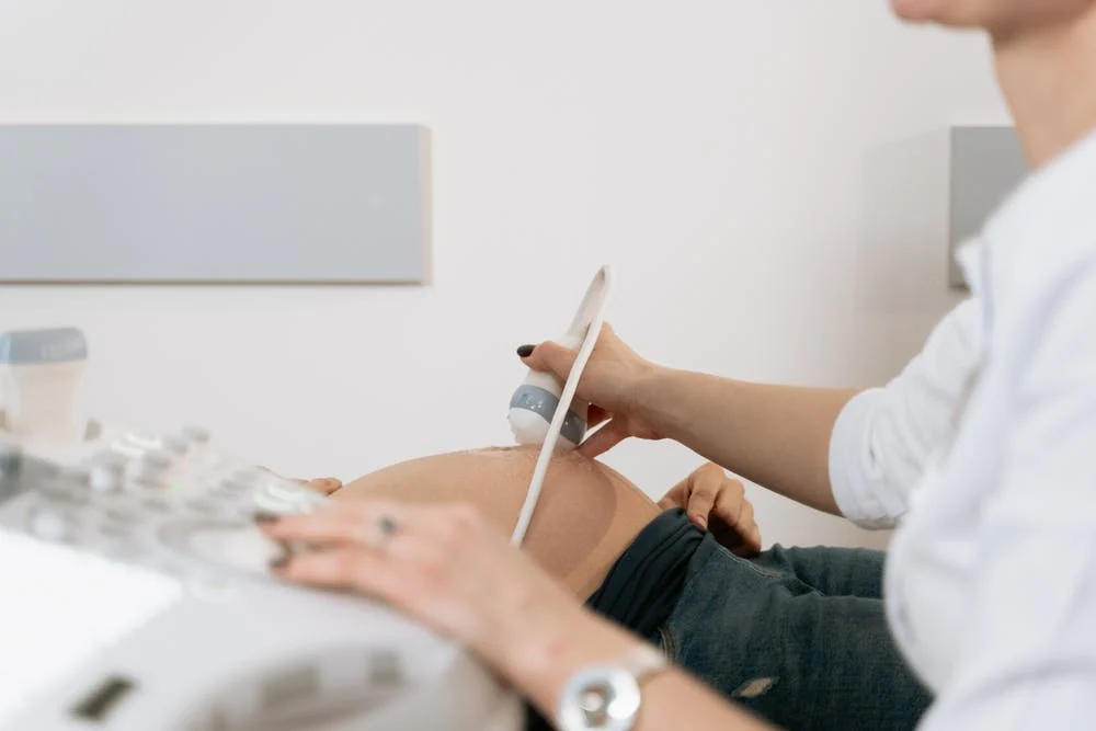
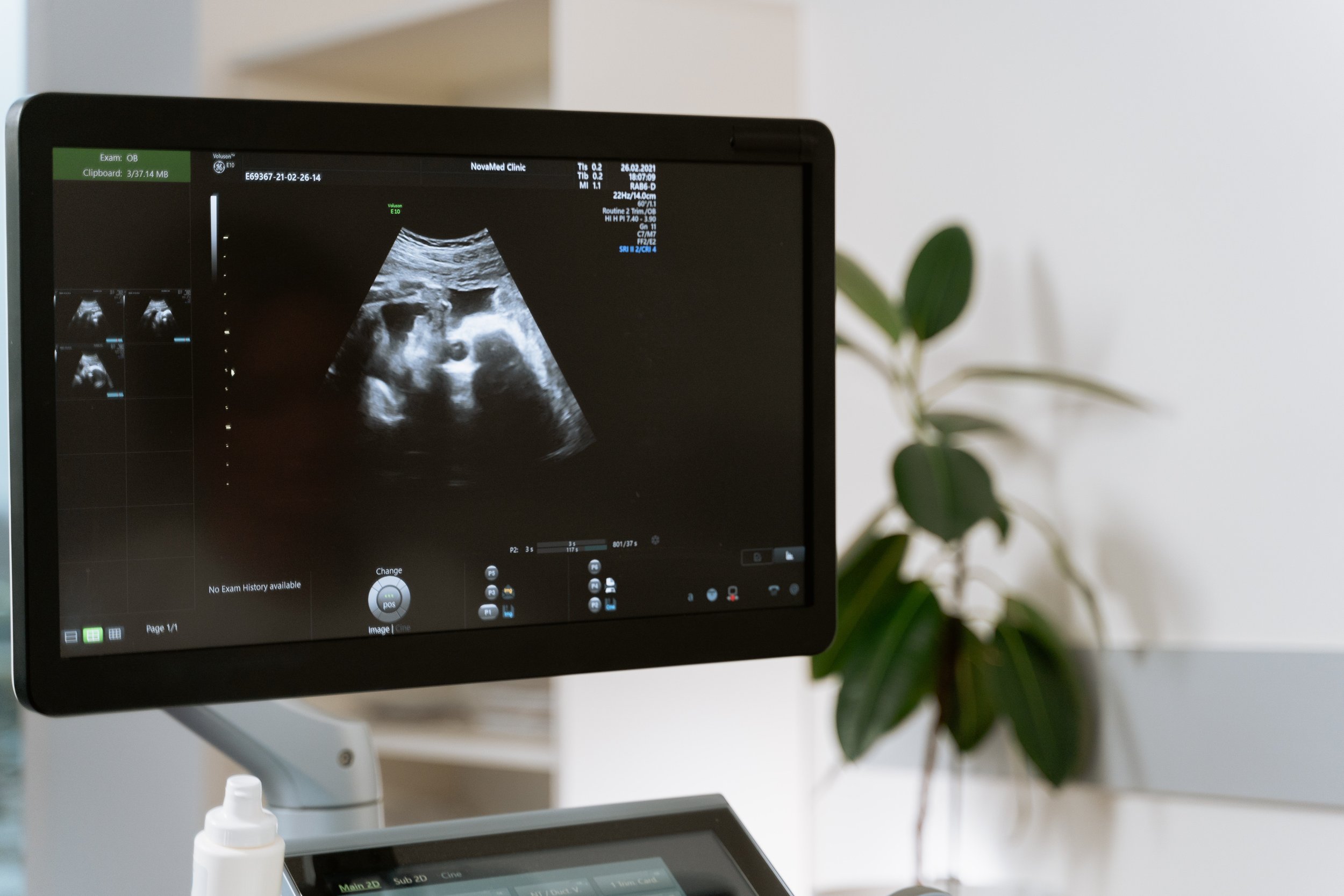
We recommend coming to a gynaecologist to register your pregnancy around 6-9 weeks of gestation. The pregnancy registering package includes an appointment with a gynaecologist, vaginal ultrasound examination, blood and urine analysis. During the first visit, the gynaecologist will assess your health, will consult and advise you about the pregnancy monitoring available in Valvekliinik, and will perform a vaginal ultrasound examination. During that ultrasound, the doctor will assess the size of the pregnancy, viability, number of fetuses, and the state of the ovaries.
We will test you for HIV, syphilis and hepatitis B. We will also determine the blood type, haemoglobin level, blood sugar level and rhesus factor. A urine test examines the presence of glucose, protein, leukocytes and bacteria and we perform a urine culture to rule out urinary tract infections.
-
The best time to perform the second-trimester screenings is between 19 and 21 weeks of gestation. The pregnancy second-trimester package includes an appointment with a gynaecologist, fetal anatomy ultrasound screening, blood and urine analysis. During the gynaecologist appointment, we assess the weight, presence of swelling, check the blood pressure, fundal height, pregnancy risks, and consult regarding any questions or complaints.
During the fetal anatomy screening, we assess the development of the fetus, the fetal heart rate, the location and structure of the placenta, and umbilical cord attachment to the placenta. We also assess the length of the pregnancy, adjust the due date, if needed. If the parent(s) wish, we check the sex of the baby.
-
The best time to perform the third-trimester screenings is between 30 and 36 weeks of gestation. The third-trimester package includes an appointment with a gynaecologist, third-trimester ultrasound screening, blood and urine analysis.
During the gynaecologist appointment, we consult regarding any questions and complaints. We assess your weight and swelling, measure blood pressure and fundal height. We take samples for HIV test and measure the haemoglobin levels, determine the blood group and rhesus factor. The third-trimester ultrasound screening is used to assess the approximate weight of the fetus, the amount of amniotic fluid, the location of the placenta, the well-being of the fetus, the posture and position of the fetus, and visualize any structural anomalies of the fetus.
Additional
-
It is possible to perform the Niptify test in the Gynaecology and Pregnancy Center of Valvekliinik. It is a non-invasive examination for the fetus, which gives the opportunity of assessing the risk of chromosomal diseases very precisely. The accuracy rate of Niptify is 99% and in some cases, it can be used to avoid unnecessary invasive procedures. The Niptify test is safe and fast, with only a blood sample taken from the mother’s vein and the test can be performed from the 10th week of pregnancy.
Niptify analyses all 23 pairs of chromosomes and identifies the four main chromosomal diseases:
Down syndrome
Edwards syndrome
Patau syndrome
Turner syndrome
The Niptify test can also identify other chromosomal deviations or random findings that can have an impact on the health of the fetus and the pregnancy. The test can also be used to give 100% exact information about the sex of the fetus. We recommend the Niptify test to all mothers who want the peace of mind that their future baby does not have four of the most common chromosomal diseases or for mothers who have performed the OSCAR test and as a result have been advised that there is a high risk of chromosomal diseases.
-
The first-trimester screening, also called the OSCAR test, is recommended for all pregnant women between 11 and 14 weeks of gestation. The OSCAR test assesses the risk of chromosomal disease and preeclampsia and monitors the anatomical development of the fetus.
The OSCAR screening is a combined test made up of two parts: a blood test and an ultrasound examination. What kind of information is received from the OSCAR test? Risk assessment of three chromosomal diseases – Down, Edwards, and Patau syndrome.
Risk assessment for preeclampsia. Preeclampsia is a disease in which the mother’s blood pressure rises in the second half of pregnancy, protein appears in the urine, changes in blood tests may occur and, in some cases, growth retardation may occur. It is possible to assess the risk of preeclampsia very accurately in the first trimester. If there is an increased risk, the woman can start prevention with aspirin. OSCAR test also concentrates on examining the fetal anatomy, as different organ systems are already visible and their development can be assessed based on the size of the pregnancy. It is possible to specify the due date.
If the OSCAR test results show a high risk of chromosomal diseases, the woman is offered additional examinations. In some cases, a fetal cell-free DNA test, called the Niptify test, may be performed but it is often advisable to perform an invasive test, such as a chorionic biopsy or amniocentesis test. Invasive tests are performed in a hospital and referenced by the doctor who performed the OSCAR test. You are welcome to OSCAR test alone or with a companion.
*The price of the OSCAR test is valid for one child, the price is doubled for twins.
-
At Valvekliinik, we perform echocardiography of the fetus, i.e. an ultrasound examination of the fetal heart. The fetal echocardiography analyses the following:
How the fetal heart works
The construction and development of the fetal heart
The blood vessels and system of the fetus
The development of the heart as a whole
We recommend coming to us for fetal echocardiography during the second trimester, more exactly between weeks 19 and 21 of gestation. We recommend this examination if previous fetal ultrasound examinations have led to suspecting issues with heart failure or arrhythmia, or if the first-trimester screening showed nuchal translucency higher than the norm.
Fetal echocardiography is also recommended if there is a history of heart failure in the family, if the mother suffers from diabetes, an auto-immune disease, or is taking anticonvulsant medicines.
*The price of the echocardiography is valid for one child, the price is doubled for twins.
-
It is possible to receive pictures of your fetus on a USB drive or have them printed out to give you something to keep from these special moments.
Our Gynecologist
-

Liina Rajasalu
GYNECOLOGIST